KEYNOTE SPEAKERS
|
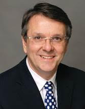
Julien DOYON
Director of the McConnell Brain Imaging Centre, The Neuro, McGill University
|
Title:Brain and Cervical Spinal Cord Contributions to Motor Learning Examined Using Functional magnetic resonance imagingAbstract:For more than 30 years, my laboratory has used psychophysical, electrophysiological and multimodal neuroimaging techniques in healthy individuals and clinical populations to investigate the behavioural determinants, neural substrates and neurophysiological mechanisms mediating the different learning phases (fast, slow, consolidation, automatization and retention) of motor skills. During this presentation, I will first review some of our work focusing on motor sequence learning (MSL) and will discuss our studies showing that the consolidation of this form of memory trace depends upon greater functional integration of the cortico-striatal system and non-rapid eye movement (N-REM) sleep spindle activity measured during the night following the initial training session. Yet despite such advances, models of motor skill learning have up until recently been incomplete because they do not account for the contribution of another important part of the central nervous system (CNS) : i.e., the cervical spinal cord (CSC). To address this knowledge gap, my group has pioneered a methodological technique that employs simultaneous brain/CSC functional magnetic resonance imaging (fMRI) during MSL in healthy individuals. In 2015 we used this new imaging method to provide the first ever evidence for local, intrinsic plasticity within the CSC, as well as connectivity changes with the sensorimotor cortex and cerebellum over the course of MSL using neuroimaging. Later, we then reported that further local functional plasticity within the CSC can be observed at different phases (fast vs. slow) of MSL using both univariate and data-driven multivariate approaches, depending on the group of muscles involved in the motor task. Finally, I will then discuss these results and highlight a few possible applications of this brain/CSC imaging technique.Biography:Julien DOYON, PhD, is the Director of the McConnell Brain Imaging Centre. He has held several leadership roles related to neuroimaging, including most recently as Scientific Director of the Unité de Neuroimagerie Fonctionnelle at the Centre de recherche, Institut universitaire de gériatrie de Montréal, Director of the Quebec Bio-Imaging Network, and codirector of the Laboratoire international de neuroimagerie et modélisation, INSERM‐ Université de Montréal. . |
|
|
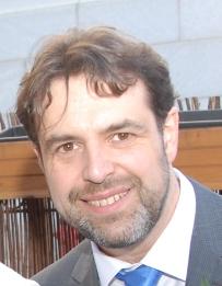
Christophe GROVA
Associate Professor, Physics / PERFORM Centre, Concordia University, Montreal, Canada
|
Title:EEG/MEG source imaging of transient and oscillatory epileptic brain activity using the maximum entropy on the mean frameworkAbstract:Accurate delineation of the epileptogenic zone (EZ) during presurgical workup of focal drug-resistant epilepsy patients can be challenging. Stereo-electroencephalography (SEEG) recordings, considered as the gold-standard for the localization of the EZ, might be the step towards mapping the seizure-onset zone (SOZ) and determining surgical candidacy. However, a successful investigation requires a strong pre-implantation hypothesis on the localization of the EZ, which can be derived from non-invasive investigations such as EEG or Magnetoencephalography (MEG) source imaging. The purpose of this talk is to introduce the Maximum Entropy on the Mean (MEM) source imaging framework, as a Bayesian approach to solve the ill-posed inverse problem of localizing the generators of EEG and MEG signals along the cortical surface. We will first review the time-domain version of MEM, which is sensitive to the spatial extent of the underlying generators, notably when localizing transient epileptic discharges. The localization accuracy of the MEM method and its ability to recover the spatial extent of the generators was quantitatively validated using SEEG (Abdallah et al Neurology 2022) or surgical cavity and postsurgical outcome as ground truth. In the second part of the talk, we will introduce the time-frequency wavelet-based extension of MEM (wavelet MEM) as a source image method of interest to localize transient oscillations, such as ictal oscillations localizing the seizure onset zone, transient high frequency oscillations and also resting state ongoing oscillations. The accuracy of wavelet-based MEM to recover oscillatory power spectra from resting state MEG data was validated using the MNI SEEG atlas of normal brain activity as ground truth (Afnan et al Neuroimage 2023).Biography:Christophe GROVA, After completing in 2002 a PhD in biomedical engineering from University of Rennes 1 (France, 1998-2002), validating multimodal image registration techniques, Dr. GROVA did a postdoctoral fellowship at the Montreal Neurological Institute (McGill University, Montreal, Canada) under the supervision of Dr. J. Gotman, studying simultaneous Electro-EncephaloGraphy (EEG)/functional Magnetic Resonance Imaging (fMRI) investigations of epileptic activity and developing expertise in EEG and MagnetoEncephaloGraphy source imaging. Recruited as assistant Professor in Biomedical Engineering and Neurology and Neurosurgery departments at McGill University in July 2008, he created the Multimodal Functional Imaging Lab, aiming at characterizing normal and pathological brain activity, especially epilepsy, combining bioelectrical neuronal activity using electrophysiology. In July 2014, he joined Concordia University as “tenure track” assistant Professor in the department of Physics of Concordia University, in the context of a strategic recruitment with PERFORM centre. PERFORM is new multimodal imaging center developed at Concordia University, dedicated to the promotion of research projects involving prevention in health and lifestyle experiences (sleep, physical exercise, nutrition). Promoted associate Professor with tenure since July 2017, his research involves combining different neuroimaging modalities to characterize brain activity, from a bio-electrical point of view (Electro-and MagnetoEncephalography) as well as from an hemodynamic perspective (functional Magnetic Resonance Imaging, Near Infra-red Spectroscopy), to study neurovascular coupling processes and ongoing resting state activity. . |
|
|
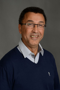
Habib BEN ALI
Scientific Director, PERFORM Centre and Professor, Department of Electrical and Computer Engineering Faculty of Engineering and Computer Science Concordia University Montreal, QC,
|
Title:Digital brain activity and pathology in Alzheimer disease: Toward a predictive physiopathologyAbstract:The objective of this research program is to better understand brain activity in healthy aging and to shed light on factors predicting conversion to neurodegenerative disease. Indeed, understanding neuronal activity, brain metabolism and patho-physiological process will enable the development of innovative computational models by combining biological and biomedical images from the basic modelling of the brain's anatomo-functional networks to models of tau protein accumulation. In such “virtual brain activity and pathology” environment, the outcome of brain disease development for individual subjects can be foreseen by simulations. Numerical simulation tools would allow prediction of the progress of the disease as well as an understanding of its causes, which remain uncertain. This computational approach is a new paradigm of the study of the patho-physiological process in healthy aging of subjects at-risk for neurodegenerative disease. This approach, referred to as “predictive physiopathology”, offer better health through prevention.Biography:Habib BEN ALI completed my PhD in Applied Mathematics and Statistics at Rennes University in 1985. He joined the French National Institute of Health and Medical Research (INSERM) in 1989. He served as Head of the Laboratory of Functional Imaging (INSERM U678 unit with over 65 members) from 2008 to 2013 and Deputy Director of the Biomedical Imaging Laboratory (with over 100 members), INSERM - The National Center for Scientific Research (CNRS) and Sorbonne University until 2015. Together with Prof. Julien DOYON, from the Université de Montréal (UdeM), he founded and became co-director of the International Laboratory of Neuroimaging and Modelisation of the INSERM-Sorbonne University and UdeM administrative bodies in 2007. He is a regular research member of the Centre de Recherche Mathématiques of UdeM since 2002 and a researcher at Centre de Recherche de l'Institut Universitaire de Gériatrie de Montréal, UdeM, since 2005. He is currently the Scientific Director for the PERFORM Centre, NSERC Canada Research Chair Tier 1 in Biomedical Imaging and Healthy Aging and Professor at the Faculty of Engineering and Computer Science at Concordia University. In the last 30 years, he has gone from starting a new neuroimaging research activity to directing a productive laboratory in France and Canada with activities that span from the development of new mathematical modeling and image analysis techniques to cognitive neuroscience and clinical applications. During this productive time in his scientific career, he published more than 300 peer reviewed papers in the best scientific journals. He is a member of national and international per review committees, executive committees and member of Advisory Board & committees. |
|
|
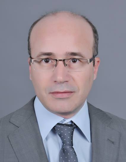
Mehrez ZRIBI
CESBIO
|
Title:Abstract:Biography:M. ZRIBI is a Director of Research in CNRS (French National Scientific Research Center) and the Head of CESBIO (Centre d’Etudes Spatiales de la BIOsphère). In 1995, he joined the Centre d’Etude des Environnements Terrestre et Planétaires Laboratory [Institut Pierre Simon Laplace/CNRS], Vélizy, France. In 2001, he joined CNRS organism to develop microwave remote sensing research applied to land surface. Since October 2008, he has been with the Centre d’Etudes Spatiales de la Biosphère, Toulouse. During the period (2008-2012), he was with the French Institute of Research for Developement to develop researches in water resources based on remote sensing in semi-arid regions. He is expert on microwave remote sensing applied to land surfaces. Mehrez ZRIBI has published actively in refereed journals with high impact factor (150 papers). He coordinated publication of 20 books about remote sensing for land surfaces. His H index (Web of Science) is equal to 48. He coordinated and participated to several research projects funded by different research programs (CNES, ANR, ESA, FP5, FP6, FP7 etc). He is associated editor for three international journals (Geophysical instrumentation, Methods and data systems (EGU), Nature/Scientific Reports and Remote Sensing/MDPI). He is senior member of IEEE Geoscience and Remote Sensing. |
|
|
Habib ZAIDI
Ph.D, FIEEE, FAIMBE, FAAPM, FIOMP, FAAIA, FBIR
|
Title:Abstract:Biography:Habib Zaidi is Chief physicist and head of the PET Instrumentation & Neuroimaging Laboratory at Geneva University Hospital and full Professor at the medical school of the University of Geneva. He is also a Professor at the University of Groningen (Netherlands), the University of Southern Denmark (Denmark) and Óbuda University (Hungary). His research is supported by the Swiss National Foundation, the European Commission, private foundations and industry (Total 10M+ US$) and centres on hybrid imaging instrumentation (PET/CT and PET/MRI), computational modelling and radiation dosimetry and deep learning. He was guest editor for 14 special issues of peer-reviewed journals and serves and serves as founding Editor-in-Chief (scientific) of the British Journal of Radiology (BJR)|Open, Deputy Editor for Medical Physics and is on the editorial board of leading journals in medical physics and medical imaging. He has been elevated to the grade of fellow of the IEEE, AIMBE, AAPM, IOMP, AAIA and the BIR. His academic accomplishments in the area of quantitative PET imaging have been well recognized by his peers since he is a recipient of many awards and distinctions among which the prestigious (100’000$) 2010 Kuwait Prize of Applied Sciences (known as the Middle Eastern Nobel Prize). Prof. Zaidi has been an invited speaker of over 170 keynote lectures and talks at an International level, has authored over 420+ peer-reviewed articles (h-index=76, >22’000+ citations) in prominent journals and is the editor of four textbooks. |
|
|
Bhiksha RAMAKRISHNAN
Carnegie Mellon University, Pittsburgh,
|
Title:
Privacy and Security Issues in Speech Processing
|
|

|
Gérard CHOLLET
Télécom-SudParis (SAMOVAR),
|
Title:
|
|
